MRI or MR-wrong
Magnetic Resonance Imaging (MRI) is a powerful diagnostic tool widely used in modern medicine, especially for musculoskeletal conditions like low back pain. However, its overuse, particularly without ruling out critical red flags, has led to an increase in unnecessary surgeries, rising healthcare costs, and patient burden. This blog post explores the impact of MRI overuse, the consequences it brings, and how healthcare professionals and patients can work together to ensure its more appropriate use.
Nhehern N. Acharya (PT)
8/27/20249 min read
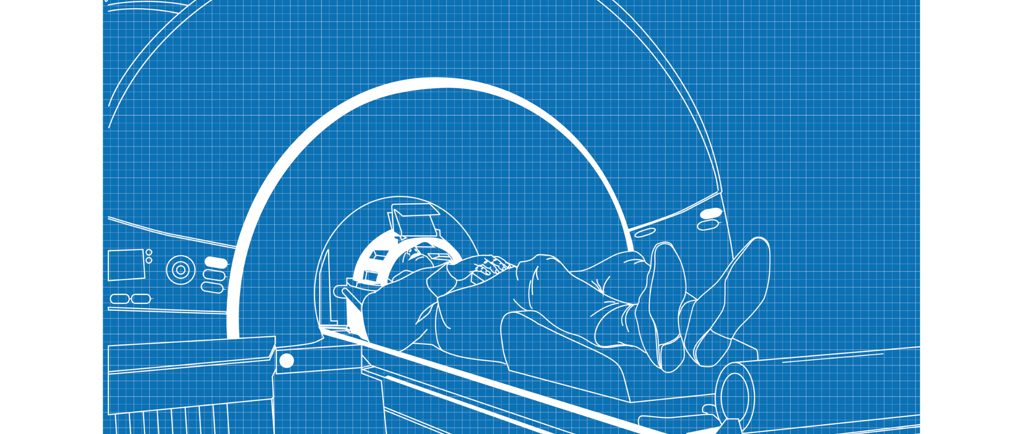
The Overuse of MRI: A Path to Unnecessary Surgeries and How to Prevent It.
Magnetic Resonance Imaging (MRI) is one of the most advanced diagnostic tools available in modern medicine, offering detailed images of soft tissues, bones, and other internal structures. For conditions involving the musculoskeletal system, particularly the spine, MRI has become a cornerstone in diagnosing various disorders. However, with great power comes great responsibility, and the overuse of MRI—especially in cases of low back pain without ruling out clinical red flags—has led to a significant increase in unnecessary surgeries, healthcare costs, and patient burden.
In this blog post, we'll explore the nuances of MRI overuse, the consequences of such practices, and how healthcare professionals and patients can work together to ensure a more judicious use of this powerful tool.
1. The Role of MRI in Modern Medicine
MRI technology has revolutionized the way we diagnose and understand musculoskeletal conditions. Unlike X-rays or CT scans, MRI uses powerful magnets and radio waves to create detailed images of the body's internal structures without exposing patients to ionizing radiation. This makes it especially valuable for diagnosing conditions involving soft tissues—like ligaments, tendons, and muscles—along with the intricate anatomy of the spine and brain.
In cases of low back pain, an MRI can reveal abnormalities such as:
Disc Herniations: Protrusions of the intervertebral discs that may impinge on nearby nerves.
Spinal Stenosis: Narrowing of the spinal canal that can compress the spinal cord or nerves.
Degenerative Disc Disease: Age-related changes in the intervertebral discs that may cause pain or stiffness.
Spondylolisthesis: Slippage of one vertebra over another, potentially leading to nerve compression.
Facet Joint Arthritis: Degenerative changes in the joints between vertebrae.
These detailed images can be incredibly valuable for pinpointing the source of a patient’s symptoms and guiding treatment decisions. However, this same detail can also be a double-edged sword.
The Appeal of MRI in Diagnosis
The allure of MRI lies in its ability to provide a clear and detailed view of the body’s internal structures, which can be reassuring for both patients and clinicians. For patients, an MRI offers the promise of clarity—a definitive answer to the question, “What’s causing my pain?” For clinicians, it provides a tool that can substantiate clinical suspicions, aiding in diagnosis and treatment planning.
However, this clarity can also lead to overconfidence in the imaging findings, potentially overshadowing the clinical context. The result? An overreliance on MRI as the ultimate diagnostic tool, sometimes at the expense of a thorough clinical evaluation.
2. The Importance of Clinical Evaluation and Ruling Out Red Flags
Before we dive into the specifics of MRI overuse, it’s important to discuss the concept of "red flags" in clinical practice. Red flags are clinical signs and symptoms that may indicate the presence of serious underlying conditions requiring immediate attention. In the context of low back pain, these red flags include:
Unexplained Weight Loss: Could suggest malignancy or systemic disease.
Severe or Progressive Neurological Deficits: May indicate spinal cord compression, cauda equina syndrome, or other serious conditions.
History of Cancer: Raises suspicion for metastatic disease, particularly in the spine.
Recent Significant Trauma: Increases the likelihood of fractures or significant ligamentous injuries.
Prolonged Use of Steroids or Immunosuppressants: Associated with an increased risk of infections and fractures.
When these red flags are present, advanced imaging like MRI is often warranted to rule out conditions such as spinal tumors, infections, or fractures. However, in the absence of these red flags, most cases of acute low back pain are self-limiting and respond well to conservative treatment.
Guidelines for Imaging in Low Back Pain
Clinical guidelines from various professional organizations, including the American College of Physicians (ACP) and the American Academy of Family Physicians (AAFP), recommend against routine imaging for low back pain in the absence of red flags. These guidelines emphasize that early imaging is unlikely to improve outcomes and may, in fact, lead to unnecessary interventions.
For instance, the ACP guidelines suggest the following approach:
History and Physical Examination: Focus on identifying red flags that may indicate a need for imaging.
Conservative Management: For patients without red flags, initial treatment should include education, self-care, physical therapy, and non-steroidal anti-inflammatory drugs (NSAIDs).
Reevaluation: If symptoms persist or worsen after 4-6 weeks of conservative management, consider imaging.
By following these guidelines, clinicians can reduce the likelihood of unnecessary imaging and interventions, ultimately improving patient outcomes and reducing healthcare costs.
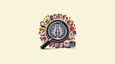
3. The Pitfalls of Misleading MRI Findings
One of the most significant challenges in using MRI as a diagnostic tool is the potential for misleading findings. Research has shown that many MRI-detected abnormalities are common even in asymptomatic individuals—those who do not have any symptoms of back pain or discomfort.
The Prevalence of "Normal Abnormalities"
Studies have consistently demonstrated that many findings on MRI, such as disc herniations or degenerative changes, are often present in people without any symptoms. For example:
Disc Herniations: A study published in The New England Journal of Medicine found that up to 76% of asymptomatic individuals aged 20 to 80 had some form of disc bulge or herniation on MRI.
Degenerative Disc Disease: Another study found that nearly 90% of individuals over the age of 60 had some degree of disc degeneration, even if they were completely asymptomatic.
Spinal Stenosis and Facet Joint Arthritis: These are common findings in the aging population and do not necessarily correlate with the presence of pain or functional limitations.
The prevalence of these "normal abnormalities" means that simply identifying them on an MRI does not necessarily explain a patient’s symptoms. Without careful correlation with the patient’s clinical history and physical examination, there is a risk of attributing symptoms to incidental findings—leading to overdiagnosis and overtreatment.
The Psychological Impact of MRI Findings
Beyond the clinical implications, MRI findings can also have a significant psychological impact on patients. The mere presence of abnormalities on imaging can lead to increased anxiety and a heightened focus on the perceived “damage” in the body. This phenomenon, sometimes referred to as "scanxiety," can exacerbate the patient’s pain experience and contribute to a cycle of chronic pain.
Patients may become overly concerned with the structural findings on their MRI, believing that their pain is directly linked to these abnormalities. This belief can lead to a sense of hopelessness and a perceived need for surgical intervention to "fix" the problem. In reality, many of these findings are benign and may not be the true source of the patient’s pain.
4. The Domino Effect: From MRI Overuse to Unnecessary Surgeries
The overuse of MRI can initiate a cascade of events that ultimately lead to unnecessary surgeries—a phenomenon often referred to as the "domino effect." This effect begins with the identification of incidental findings on MRI and can progress through several stages:
Detection of Incidental Findings- As previously discussed, MRIs often reveal abnormalities that may not be related to the patient’s symptoms. These findings, while common, can create a sense of urgency or concern among both patients and clinicians, leading to further investigation and intervention.
Increased Anxiety and Focus on Abnormalities- Once abnormalities are identified on MRI, patients and clinicians may become fixated on these findings. The patient may interpret the results as evidence of a serious underlying problem, while the clinician may feel compelled to act on the findings, even if they are incidental.
Referral to Specialists- The next step in the domino effect is often a referral to a specialist, such as an orthopedic surgeon or neurosurgeon. The specialist may recommend additional imaging or diagnostic tests to further evaluate the abnormalities identified on MRI. In some cases, the specialist may suggest surgical intervention as a way to address the perceived problem.
Surgical Interventions- The culmination of the domino effect is often surgical intervention. Surgeries such as spinal fusion, discectomy, or laminectomy may be performed to "correct" the abnormalities identified on MRI. However, these surgeries may not address the true source of the patient’s pain, particularly if the MRI findings were incidental.
Potential for Unnecessary Procedures- The consequence of this domino effect is a significant increase in the number of unnecessary surgeries performed each year. These surgeries carry inherent risks, including:
Infection: A common complication of any surgical procedure.
Nerve Damage: Particularly in spinal surgeries, there is a risk of damage to the nerves, which can result in chronic pain, weakness, or loss of function.
Prolonged Recovery: Spinal surgeries often require lengthy recovery periods, during which patients may experience significant discomfort and limitations in their daily activities.
Failure to Relieve Symptoms: Perhaps the most concerning outcome is the possibility that the surgery will not alleviate the patient’s symptoms. In some cases, patients may even experience worsening of their pain after surgery.
The overuse of MRI and the subsequent increase in unnecessary surgeries contribute to a growing burden on the healthcare system. It also raises ethical concerns about the potential harm to patients who undergo invasive procedures that may not be necessary.
5. Emphasizing Conservative Management and Clinical Judgment
Given the risks associated with the overuse of MRI and the potential for unnecessary surgeries, it is crucial to emphasize the importance of conservative management and clinical judgment in the treatment of low back pain and other musculoskeletal conditions.
The Value of Conservative Management
Conservative management refers to non-surgical approaches to treating musculoskeletal conditions. This may include:
Physical Therapy: Tailored exercises and manual therapy techniques can help alleviate pain, improve mobility, and strengthen the muscles supporting the spine.
Medication: Non-steroidal anti-inflammatory drugs (NSAIDs) and muscle relaxants may be used to manage pain and inflammation.
Education and Self-Care: Patients should be educated about their condition, including the importance of staying active and avoiding prolonged bed rest. Self-care strategies, such as hot/cold therapy and posture correction, can also be effective.
Cognitive Behavioral Therapy (CBT): In cases of chronic pain, CBT can help patients manage the psychological aspects of pain, such as anxiety and depression.
By focusing on conservative management, clinicians can help patients avoid the risks associated with unnecessary imaging and surgery. In many cases, conservative treatment is sufficient to alleviate symptoms and improve function, allowing patients to return to their normal activities without the need for invasive procedures.
The Importance of Clinical Judgment
Clinical judgment is the cornerstone of effective patient care. It involves integrating the patient’s history, physical examination findings, and any relevant imaging or diagnostic tests to arrive at a diagnosis and treatment plan. While MRI is a valuable tool, it should not be used as a substitute for clinical judgment.
Healthcare professionals should be cautious in interpreting MRI findings and avoid relying solely on imaging to make treatment decisions. Instead, they should consider the broader clinical context, including the patient’s symptoms, functional limitations, and response to conservative treatment.
Shared Decision-Making
Shared decision-making is a collaborative process in which healthcare providers and patients work together to make informed decisions about treatment options. This approach emphasizes the importance of patient education and engagement, allowing patients to understand the potential risks and benefits of different treatment options.
When discussing MRI findings and potential treatment options, clinicians should take the time to explain the limitations of imaging and the potential for incidental findings. They should also discuss the potential risks and benefits of surgery, as well as the likelihood of achieving the desired outcomes with conservative management.
By involving patients in the decision-making process, clinicians can help them make informed choices that align with their values and preferences. This approach can reduce the likelihood of unnecessary surgeries and improve patient satisfaction with their care.
Conclusion
The overuse of MRI in the absence of clinical red flags has led to a significant increase in unnecessary surgeries, healthcare costs, and patient burden. While MRI is a powerful diagnostic tool, it should be used judiciously and in conjunction with a thorough clinical evaluation.
By emphasizing the importance of conservative management, clinical judgment, and shared decision-making, healthcare professionals can help patients avoid the risks associated with unnecessary imaging and surgery. Ultimately, the goal is to provide high-quality, patient-centered care that improves outcomes and enhances the patient’s quality of life.
As we continue to advance in the field of diagnostic imaging, it is essential to remember that more is not always better. Thoughtful and responsible use of MRI, guided by clinical expertise and patient-centered care, is the key to ensuring the best possible outcomes for patients.


References
Saini R, Sharma A, Dave MB. Clinical Reporting of Magnetic Resonance Imaging, the Way Forward for Patients With Lumbar Disc Herniation: A Prospective Correlational Study. Cureus. 2022;14(7):e27232. Published 2022 Jul 25. doi:10.7759/cureus.27232
Jacobs JC, Jarvik JG, Chou R, et al. Observational Study of the Downstream Consequences of Inappropriate MRI of the Lumbar Spine. J Gen Intern Med. 2020;35(12):3605-3612. doi:10.1007/s11606-020-06181-7
Brinjikji W, Luetmer PH, Comstock B, et al. Systematic literature review of imaging features of spinal degeneration in asymptomatic populations. AJNR Am J Neuroradiol. 2015;36(4):811-816. doi:10.3174/ajnr.A4173
WORKING HOURS
Monday - Saturday 8:00 AM - 8:00 PM
Sunday 9:00 AM - 5:00 PM
ADDRESS
Kairos Physio, F3 First floor, near Basillios Gym, St. Inez Rd., St. Inez, Panjim- Goa 403001.
CONTACT
+91 7259527374
kairosphysio@gmail.com